Muscle Echo Intensity and Healthy Aging: What Ultrasound Can Reveal
Muscle echo intensity may reflect changes in muscle composition, but it is not a diagnosis. Learn what ultrasound shows and how to interpret the evidence.
Table of Contents
DISCLAIMER
This article is for informational purposes only and does not constitute medical advice. The statements in this article have not been evaluated by the FDA. The information presented is based on published research and should not be used as a substitute for professional medical guidance. Consult your physician before starting any supplement or health protocol.
Two people can have similar thigh size and very different strength. One reason is that muscle quantity is not the same as muscle quality. Muscle echo intensity in healthy aging is an ultrasound-based research measure that attempts to capture part of that difference by analyzing how bright or dark muscle tissue appears.
The idea is appealing: ultrasound is portable, radiation-free, and relatively inexpensive. But brightness is not a direct photograph of fat or fibrosis. It is a signal shaped by tissue composition, machine settings, probe angle, depth, and analysis choices.
What Echo Intensity Measures
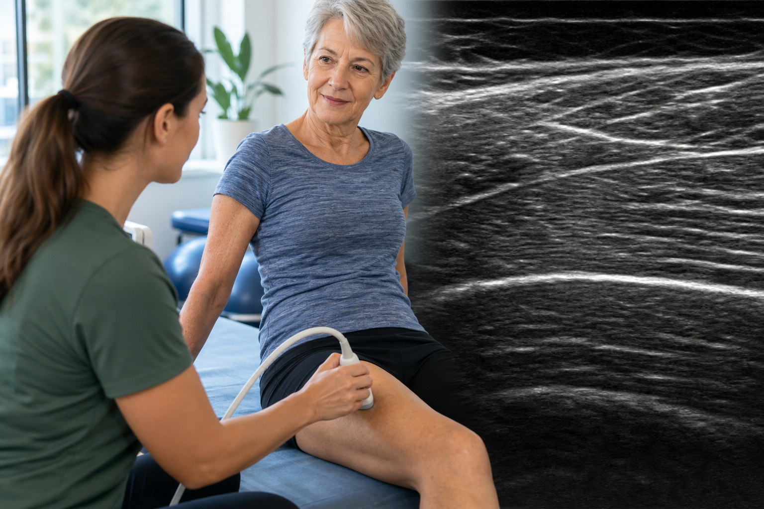
Ultrasound sends sound waves into tissue and records returning echoes. Healthy contractile muscle often appears relatively dark, crossed by brighter lines from connective tissue. When more sound is reflected, the image may look brighter and its grayscale value rises.
Higher echo intensity has been associated with intramuscular fat, connective tissue, and other non-contractile changes. Those components can influence force production and metabolic health. Yet ultrasound cannot cleanly separate them without support from MRI, CT, biopsy, or other methods.
The measure is usually calculated inside a selected region of interest. That seemingly simple choice matters: including fascia, a blood vessel, or a shadow can shift the result. Comparing raw numbers from different machines is therefore risky.
Why Muscle Quality Changes With Age
Aging muscle can lose fibers, especially fast motor units, while surviving fibers remodel. Fat may accumulate between and within muscles. Connective tissue can increase, and the architecture of fascicles can change. Inactivity, insulin resistance, inflammation, nerve injury, and illness influence the pattern.
These changes help explain why strength can decline faster than muscle size. The nervous system must recruit fibers effectively, the fibers must transmit force through connective tissue, and joints must move without excessive pain. Echo intensity observes only one slice of that system.
Hydration and recent activity can also affect tissue appearance. A hard exercise session may temporarily change fluid distribution. Standardized rest, body position, probe pressure, and anatomical landmarks are essential in research.
What Studies Have Found
Cross-sectional studies often find higher echo intensity in older groups and associations with lower strength, slower walking, or poorer chair-rise performance. Some analyses report that echo intensity explains functional variation beyond muscle thickness alone.
Longitudinal evidence is less abundant. An association at one time point cannot show whether brighter muscle caused weakness or whether inactivity and illness changed both. Different muscles may also tell different stories: quadriceps imaging is common, but it does not represent the entire body.
Training studies are mixed. Resistance exercise can increase strength and muscle thickness, while echo-intensity changes may be smaller or depend on baseline status. That is not a failure; improved neural recruitment and fiber function can matter even without a dramatic grayscale shift.
How It Fits With Other Measures
Clinical assessment of possible sarcopenia generally begins with strength, such as grip or chair stands, then considers muscle quantity and physical performance. Ultrasound could complement this process where DXA or CT is unavailable, but universal cut points are not yet established.
Muscle thickness estimates quantity, pennation angle describes architecture, and echo intensity suggests composition. Functional measures—walking speed, stair power, balance, and lifting capacity—show what the person can actually do. The most useful picture combines rather than substitutes.
For individuals, progress should not be reduced to image brightness. A person who stands more easily, carries groceries, and recovers balance has achieved meaningful improvement even if a research metric moves modestly.
Practical Perspective
Progressive resistance training provides the central stimulus for maintaining muscle capacity. Adequate protein and energy support adaptation, while aerobic activity helps metabolic and vascular health. Programs should be scaled for health, experience, and recovery.
Rapid unexplained weakness, substantial muscle wasting, repeated falls, or one-sided change deserves medical evaluation. An ultrasound image obtained in a fitness setting cannot rule out neurological, endocrine, inflammatory, or orthopedic causes.
The Bottom Line
Muscle echo intensity offers a useful research window into age-related muscle composition, particularly when paired with thickness, strength, and performance. It is not a stand-alone diagnosis or a consumer score to optimize; function remains the outcome that matters most.
Frequently Asked Questions
What does high muscle echo intensity mean?
Is muscle ultrasound used to diagnose sarcopenia?
Can training change echo intensity?
Sources
Stay Updated on Longevity Science
Weekly research digests. No spam, unsubscribe anytime.
Related Articles
Arterial Calcification and Vascular Aging: Reading the Risk Signals
Arterial calcification can reveal accumulated vascular risk. Learn what calcium scores mean, why calcification develops, and where testing has limits.
10 min read
Arterial Stiffness and Vascular Age: A Longevity Evidence Guide
Understand arterial stiffness vascular age longevity links, key studies, measurement limits, and evidence-based lifestyle implications for healthy aging.
11 min read
Arterial Stiffness and Vascular Aging: What the Evidence Shows
Explore arterial stiffness vascular aging, how vessels change over time, what pulse wave velocity studies show, and practical ways evidence may inform prevention.
10 min read